Atomic Force Microscope (AFM)
Atomic Force Microscope
Operating
Model: Dimension Icon
Function: Accurately observe the 3D topography of micro-regions (nanometer and submicron scales) on the sample surface; meanwhile, it can scan the electric field/magnetic field distribution, surface potential and other properties of the sample surface.
Engineer: Shen / +86-21- 34207734-8010 / shenyunliang@1
Location: East District Test Area III,West District Test Area I
Equipment ID: ETE3AFM01、WT1BAFM01
  • Basic Equipment Information
  • Operating Principle
  • Typical Application Case
Main Application
  • 1. Observation and measurement of 3D topography in micro-regions on sample surface (nanoscale).
  • 2. Scanning of electric field/magnetic field distribution on sample surface (EFM/MFM).
  • 3. Nanoscale measurement of surface potential (AM-KPFM).
  • 4. Piezoelectric property testing of material micro-regions (PFM).
  • 5. Nanomanipulation realized by atomic probe and automatic stage (manipulation of nanoscale materials and structures).
  • 6. Measurement modes: Smart mode, Contact mode, Tapping mode, etc.


Process/Testing Capabilities

  • XY scanning range: 90 μm × 90 μm; Z scanning range: 10 μm.
  • Equipped with Contact Mode, Tapping Mode, Phase Imaging Mode, Piezoresponse Force Microscopy Mode, and Smart Scanning Mode.


Technical Specifications

  • Intelligent automatic tip approaching method.
  • 210 mm vacuum adsorption fully automatic sample stage with programmable control.
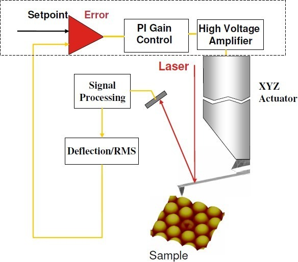
The working principle of the atomic force microscope (AFM) is based on the interatomic interaction force. When an ultra-sharp probe approaches the sample surface, weak interaction forces such as van der Waals forces are generated between the atoms of the probe and the sample surface. As the probe scans over the sample surface, the topographic information of the sample surface is acquired by detecting changes in these forces.The system takes the amplitude of the cantilever as the feedback signal. At the start of scanning, the cantilever amplitude is equal to the threshold value. When the probe scans across variations in the sample surface topography, the amplitude changes, causing the detected signal to deviate from the threshold and generate an error signal. The system eliminates the error signal through a PID controller, which drives the movement of the scanner tube, thereby recording the surface topography of the sample. The overall AFM system is shown in the figure below.

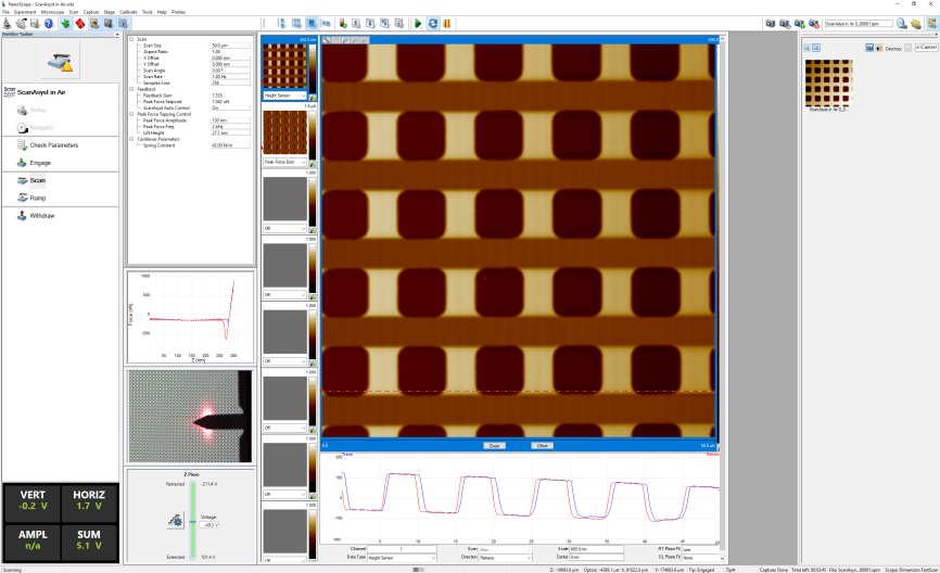
Provides an intelligent scanning mode: it adopts picoNewton-level force feedback with force curves generated by sinusoidally driven piezoelectric ceramic tubes for surface imaging, and the force curve frequency is ≥ 2000 Hz.Only the scanning range needs to be selected; the system can automatically adjust the contact force, circuit gain, scanning speed and scanner tube range during scanning.

1. Sample size shall be less than 8 inches; excessive viscosity on the sample surface is prohibited.
2. The upper and lower surfaces of the sample shall be flat and clean, free from oil stains, dust and other contaminants.
1. The sample size shall be less than 8 inches, and excessive viscosity on the sample surface is not allowed.
2. The upper and lower surfaces of the sample shall be flat and clean, free of oil stains, dust and other contaminants.
3. Keep the back side of the sample clean and close the software after use.
4. Do not touch the equipment or operating computer during operation, so as to avoid equipment shutdown or malfunction caused by accidental contact; in case of equipment alarm, please notify the corresponding equipment engineer immediately.
The list below shows FAQs (click a question to view the answer). If your question is not listed, you can leave a message using the link.
FAQs
  • 01
    原子力显微镜的扫描速度对测量结果有什么影响?

    扫描速度过快可能会导致图像失真和分辨率降低,因此需要根据样品的性质和测量要求选择合适的扫描速度。

Message Inquiry
If your question is not listed above, please email us at aemd@sjtu.edu.cn or click the button below to submit an inquiry.
Note: "@1" represents "@sjtu.edu.cn"
×